1 Diagram of the human heart. The image depicts the different cavities... Download Scientific
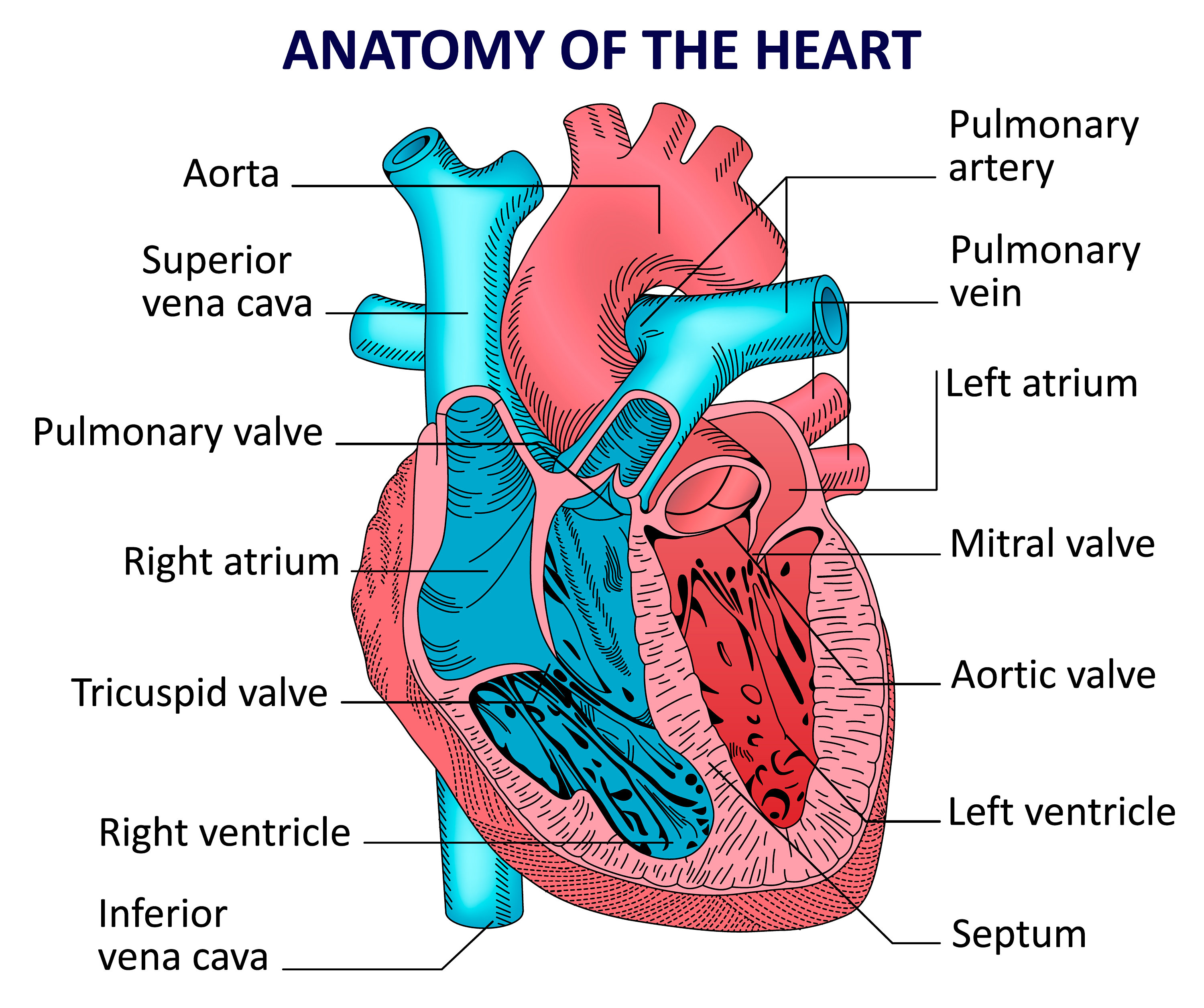
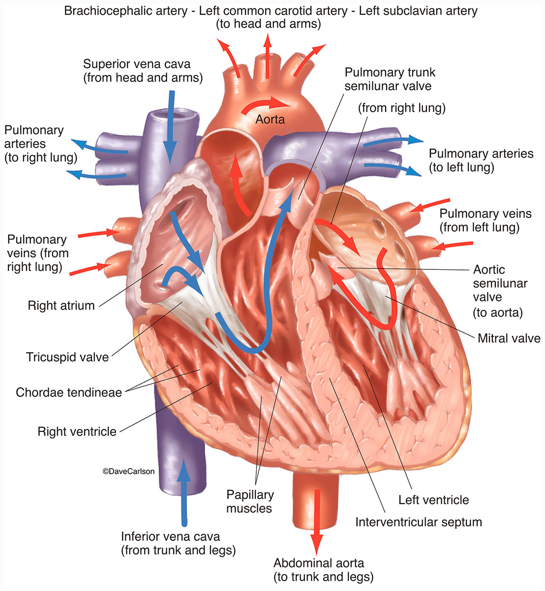
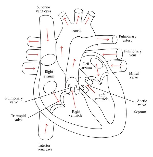
Every heart diagram labeledwill clearly show these valves. These valves allow blood flow in one direction only. Different valves perform different functions. Tricuspid valve is located between the right ventricle of your heart and the right atrium, and allows the blood to move from the right atrium to the right ventricle.

Basic Anatomy of the Human Heart Cardiology Associates of Michigan Michigan's Best Heart Doctors
A labelled diagram of the human heart. This is a featured picture on the English language Wikipedia ( Featured pictures) and is considered one of the finest images. See its nomination here. If you think this file should be featured on Wikimedia Commons as well, feel free to nominate it.

Anatomy and Physiology Heart Anatomy
Higher; Structure and function of the heart The structure of the heart. In this Higher Human Biology revision guide, you will learn in detail that cardiac output is a measure of the rate of blood.
Human heart anatomy. Vector diagram Etsy
The heart is a muscular organ that serves to collect deoxygenated blood from all parts of the body, carries it to the lungs to be oxygenated and release carbon dioxide. Then, it transports the oxygenated blood from the lungs and distributes it to all the body parts. The heart pumps around 7,200 litres of blood in a day throughout the body.; The heart is situated at the centre of the chest and.
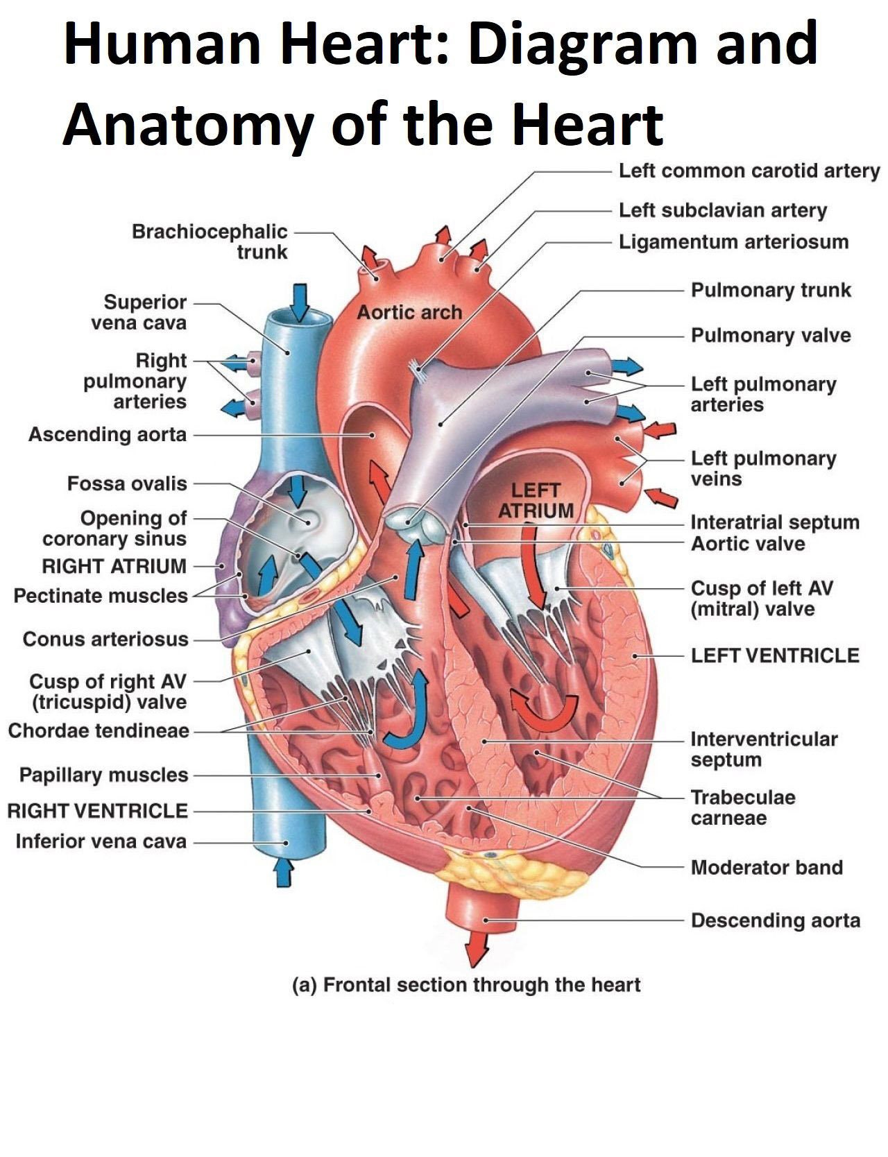
Diagram and Anatomy of the Heart Poster Etsy
Your heart is the primary organ of your circulatory system. It pumps blood throughout your body, controls your heart rate and maintains blood pressure. Your heart is a bit like a house. It has walls, rooms, doors, plumbing and an electrical system. All the parts of your heart work together to keep blood flowing and send nutrients to your other.
Human Heart Front View Interior Carlson Stock Art
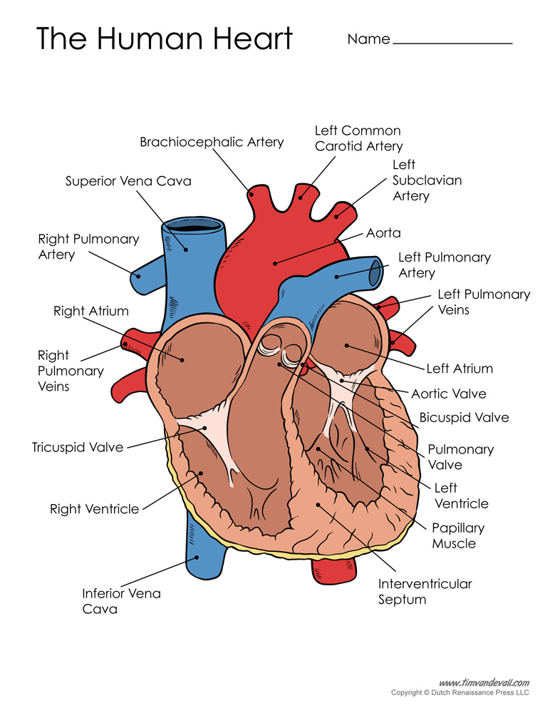
A heart is labelled as it would appear in a chest, so the left side of an image represents the right side of the heart (and vice versa). Diagram of a Human Heart. Heart Structure. The structure of the human heart includes the following key components: Chambers. Two atria (singular = atrium) - smaller chambers near top of heart that collect.
Human Heart Diagram Tim's Printables
Cardiomyopathy is when the heart muscle becomes enlarged, thick, or rigid. As cardiomyopathy worsens, the heart becomes weaker and is less able to pump blood through the body and maintain a normal electrical rhythm.
Heart Diagram with Labels and Detailed Explanation
The average human heart weighs between 6 and 11 ounces. The muscle is strong enough to pump up to 2,000 gallons — as much as a fire department's tanker truck — of blood through one's body.

Heart Disease Definition, Causes, Research
Anatomical structures were labelled according to the actual Terminologia Anatomica. Anatomy of the human heart and coronaries: how to visualize anatomic structures This tool provides access to several medical illustrations, allowing the user to interactively discover heart anatomy.

Heart Anatomy Labeled Diagram, Structures, Blood Flow, Function of Cardiac System — EZmed
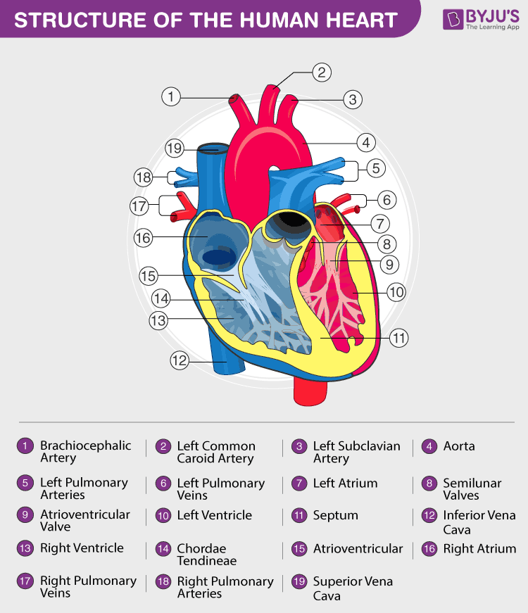
Pericardium Human heart is covered by a double layered structure which is known as pericardium. The outer layer is associated with the major blood vessels whereas the inner layer is attached to the cardiac muscles. These layers are separated by a pericardial fluid. This covering is like a membrane which holds all the parts of the heart. Chambers
Human Heart Anatomy and Functions Location and Chambers
Size of this PNG preview of this SVG file: 611 × 600 pixels 244 × 240 pixels 489 × 480 pixels 782 × 768 pixels 1,043 × 1,024 pixels 2,086 × 2,048 pixels 663 × 651 pixels. (SVG file, nominally 663 × 651 pixels, file size: 93 KB) PNG, JPG, GIF. Licensing. I, the copyright holder of this work, hereby publish it under the following licenses.

External Structure Of Heart Anatomy Diagram
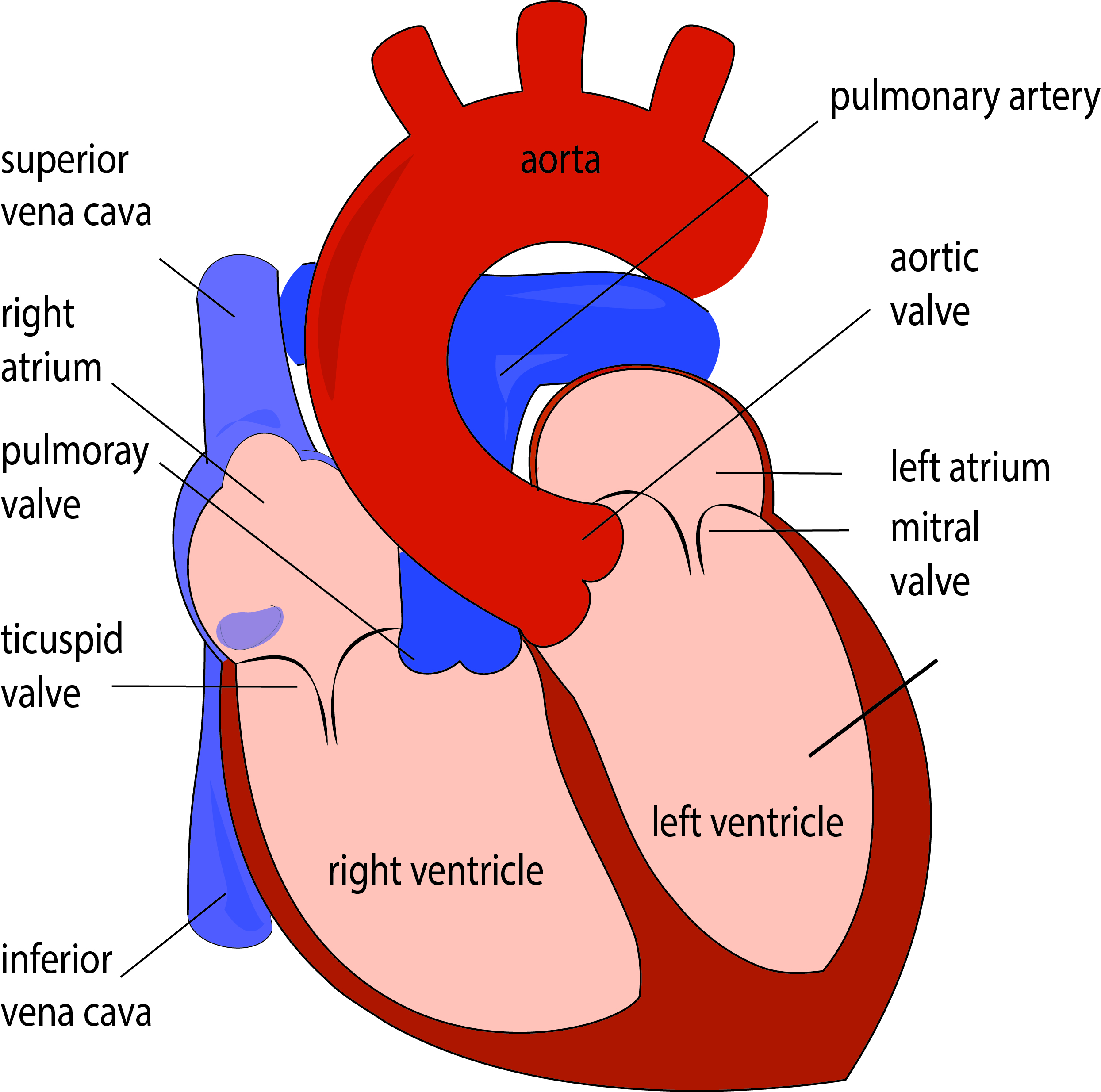
Well-Labelled Diagram of Heart The heart is made up of four chambers: The upper two chambers of the heart are called auricles. The lower two chambers of the heart are called ventricles. The heart wall is made up of three layers: The outer layer of the heart wall is called epicardium. The middle layer of the heart wall is called myocardium.

Human Heart Diagram Labeled
The heart is a vital organ of the body; therefore, minute dysfunctions or abnormalities in the heart may have drastic consequences on human health. Typically, the heart is the size of an adult.

Heart Anatomy chambers, valves and vessels Anatomy & Physiology
The human heart is primarily comprised of four chambers. The two upper chambers are called the atria, the remaining two lower chambers are the ventricles. The right and left sides of the heart are separated by a muscle called the "septum.". Both sides work together to efficiently circulate the blood.
On Heart Kardiohirurgija.rs
The heart is a muscular organ that pumps blood around the body by circulating it through the circulatory/vascular system. It is found in the middle mediastinum, wrapped in a two-layered serous sac called the pericardium.
Show me a diagram of the human heart? Here are a bunch! Interactive Biology, with Leslie Samuel
Practise Labelling the Human Heart Diagram Introduction to the Human Heart The human heart is one of the most important organs responsible for sustaining life. It is a muscular organ with four chambers. The size of the heart is the size of about a clenched fist.